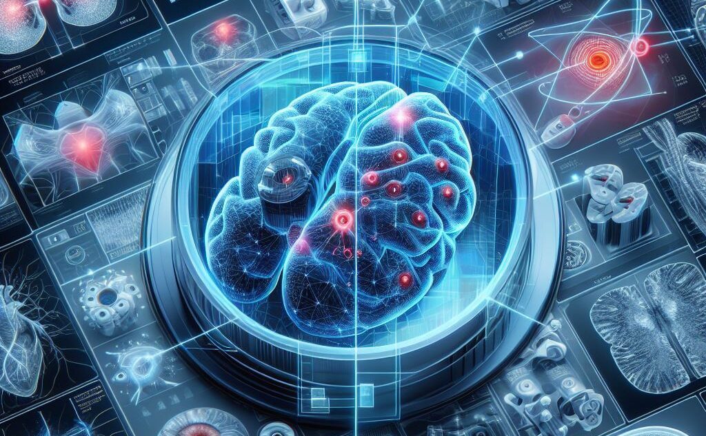
Welcome to the forefront of healthcare innovation, where the convergence of medical imaging and deep learning is reshaping the landscape of diagnosis.
Medical imaging serves as a cornerstone in modern healthcare, offering invaluable insights into the inner workings of the human body. From detecting tumors to assessing organ function, medical imaging plays a pivotal role in diagnosing diseases and guiding treatment decisions. Through techniques such as X-rays, MRIs, CT scans, and ultrasounds, clinicians gain a window into the intricate details of anatomical structures and physiological processes, enabling them to identify abnormalities, monitor disease progression, and evaluate treatment responses with unprecedented precision.
The Revolutionary Impact of Deep Learning. Enter deep learning, a subset of artificial intelligence that mimics the human brain’s neural networks to process and interpret complex data. In recent years, deep learning has emerged as a game-changer in medical imaging analysis, revolutionizing the way clinicians interpret diagnostic images and make clinical decisions. By harnessing the power of deep learning algorithms, healthcare professionals can unlock hidden insights within medical images, uncover subtle patterns indicative of diseases, and achieve diagnostic accuracy previously thought unattainable.
Deep Learning in Medical Imaging
Convolutional Neural Networks (CNNs), also known as ConvNets, are specialized deep learning architectures designed for tasks requiring object recognition from images. With their remarkable ability to autonomously extract features from data, CNNs have become indispensable in various applications, including autonomous vehicles, security camera systems, and medical imaging. Here’s an overview of the key points about CNNs:
Purpose and Applications: CNNs excel in scenarios where object recognition from images is paramount. They play crucial roles in identifying pedestrians, traffic signs, and obstacles in autonomous vehicles, detecting intruders or unusual activities in security camera systems, and diagnosing diseases from medical imaging such as X-rays, MRIs, and other scans. Their capacity to autonomously extract features at scale bypasses the need for manual feature engineering, making them efficient and versatile.
Key Features of CNNs: CNNs possess several key features that contribute to their effectiveness. They exhibit translation-invariance, meaning they can identify and extract patterns regardless of variations in position, orientation, scale, or translation. Convolutional layers enable CNNs to learn hierarchical features by applying convolution operations, allowing them to capture complex patterns. Pre-trained architectures like VGG-16, ResNet50, and EfficientNet achieve top-tier performance and can be fine-tuned for new tasks with minimal data, further enhancing their versatility and applicability.
Inspiration from the Human Visual System: CNNs draw inspiration from the layered architecture of the human visual cortex, mimicking its hierarchical structure and local connectivity. Much like the visual cortex, CNNs exhibit hierarchical organization, with early layers detecting simple features and deeper layers recognizing complex patterns. Local connectivity ensures that neurons are connected to local regions, similar to the visual cortex, while pooling layers summarize local features, making CNNs robust to translations.
Achievements and Future Research: CNN architectures have evolved significantly over the years, with milestones such as AlexNet, ResNet, and SENet pushing the boundaries of image classification. Researchers continue to explore novel architectures and techniques to further enhance CNN performance. Transfer learning and fine-tuning, where pre-trained CNNs are adapted to specific tasks with limited data, have accelerated model development and deployment. Moreover, CNNs extend beyond image classification, finding applications in natural language processing, time series analysis, and speech recognition, showcasing their versatility and potential for innovation in diverse domains.
Introduced by Ian Goodfellow and colleagues in 2014, Generative Adversarial Networks (GANs) represent a ground-breaking approach to modelling and generating data in an unsupervised manner. The acronym “GAN” stands for Generative Adversarial Networks, capturing the essence of the adversarial relationship between two neural networks within the framework.
Transformative Results
1. Convolutional Neural Networks (CNNs) in Medical Image Understanding
Deep learning, particularly Convolutional Neural Networks (CNNs), has made remarkable strides in medical imaging. These networks have outperformed human experts in various image understanding tasks. Here’s an overview:
Medical imaging plays a critical role in modern healthcare, aiding clinicians in diagnosis, treatment, and monitoring of diseases. Traditional image analysis methods often rely on manual feature extraction, which can be time-consuming and prone to human error.
Machine learning (ML) techniques, including SVMs, decision trees, and random forests, have been applied to medical image analysis. However, they require handcrafted features and expert knowledge. CNNs automatically learn hierarchical features from raw data, making them ideal for medical image analysis. CNNs have excelled in tasks like image classification, segmentation, localization, and detection.
High Diagnostic Accuracy Achieved by CNNs
Breast Cancer Diagnosis:
CNNs have demonstrated impressive results in breast cancer diagnosis.
National Library of Medicine
Organ Segmentation:
CNNs excel in segmenting organs from medical images.
National Library of Medicine
2.Generative Adversarial Networks (GANs)
Generative Adversarial Networks (GANs) represent another frontier in deep learning, with applications ranging from image synthesis to anomaly detection in medical imaging. GANs consist of two neural networks—the generator and the discriminator—engaged in a competitive game, where the generator learns to produce realistic synthetic images, while the discriminator learns to distinguish between real and synthetic images.
Applications in Medical Imaging:
-
- Image Synthesis: GANs can generate synthetic medical images that closely resemble real patient data, thereby augmenting scarce datasets and improving the robustness of deep learning models.
-
- Anomaly Detection: GANs can detect anomalies or outliers in medical images by identifying deviations from normal patterns, enabling early diagnosis of diseases or abnormalities.
In essence, deep learning, fueled by innovations such as CNNs and GANs, holds immense promise in revolutionizing medical imaging analysis, paving the way for more accurate, efficient, and personalized healthcare solutions. Let’s delve deeper into the remarkable results achieved by deep learning algorithms in various medical imaging tasks.
3. GANs for Data Augmentation
-
- Labeled medical imaging data is scarce and expensive to generate.
-
- Deep learning models require large amounts of diverse data for robustness and generalization.
-
- GANs offer a novel approach to data augmentation.
-
- Researchers have used CycleGAN to transform contrast CT images into non-contrast images, augmenting training data.
Benefits of GAN-based Augmentation:
-
- Enhances diagnostic accuracy.
-
- Streamlines workflow efficiency.
-
- Expands access to expert-level image analysis.
Reduces manual segmentation effort and cost in CT imaging.
deep learning, especially CNNs and GANs, has revolutionized medical imaging. These advancements hold immense promise for improving patient outcomes and healthcare efficiency.
How GANs Work:
GANs comprise two neural networks: the generator and the discriminator. The generator is responsible for creating new data samples, such as images, from random noise, while the discriminator distinguishes between real data and generated data. Through a minimax game, the generator strives to produce realistic data to deceive the discriminator, while the discriminator learns to differentiate between real and fake data. Over time, this adversarial process leads to the refinement of both networks, with the generator improving its ability to generate convincing samples.
Applications of GANs:
The versatility of GANs extends across a wide range of applications:
-
- Image Generation: GANs can produce high-quality images that closely resemble real photographs.
-
- Super-Resolution: Enhancing image resolution while preserving realism.
-
- Semantic Segmentation: Creating pixel-wise segmentations in images for tasks like object detection.
-
- Image Deblurring: Removing blur from images to enhance clarity.
-
- Text-to-Image Generation: Generating images from textual descriptions.
-
- Steganography: Concealing information within images for secure communication.
-
- Speech Recognition, Object Detection, and more.
GAN Variants and Achievements:
Researchers have developed various GAN variants to address specific challenges and enhance performance. Some notable variants include:
-
- Deep Convolutional GANs (DCGANs): Improving image quality using convolutional neural networks.
-
- Conditional GANs (cGANs): Generating data conditioned on specific attributes or labels.
-
- CycleGANs: Translating images from one domain to another, enabling tasks like style transfer.
-
- StyleGANs: Offering control over image styles and features for artistic manipulation.
GANs have achieved remarkable results across diverse domains, including medical imaging, art generation, and natural language processing, showcasing their potential for innovation and creativity.
Challenges and Future Research:
Despite their successes, GANs pose several challenges and avenues for future research:
-
- Mode Collapse: GANs may suffer from limited diversity in generated samples.
-
- Training Stability: Ensuring stable training dynamics for both the generator and discriminator.
-
- Interpretable Latent Space: Understanding and interpreting the learned representations in the latent space.
-
- Ethical Considerations: Addressing biases and fairness issues in GAN-generated data.
-
- Hybrid Models: Exploring opportunities to combine GANs with other architectures for enhanced performance and capabilities.
As researchers continue to tackle these challenges and push the boundaries of GAN technology, the future holds immense promise for further advancements in data generation and manipulation
Exploring Other Deep Learning Architectures
beyond Convolutional Neural Networks (CNNs) and explore other architectures, along with their applications in medical imaging. We’ll also discuss attention mechanisms and the crucial role they play in enhancing model interpretability. Finally, we’ll touch upon Explainable AI (XAI) methods and their significance in making deep learning models more transparent and interpretable.
Recurrent Neural Networks (RNNs) in Medical Imaging
Understanding RNNs
Recurrent Neural Networks (RNNs) are specialized neural network architectures designed for processing sequential data or time-series data. Unlike feedforward networks, RNNs have feedback connections that allow them to maintain an internal state and process data sequentially. RNNs excel at recognizing temporal dependencies and can predict the next likely data point in a sequence.
Applications of RNNs in Medical Imaging
Language Models: RNNs are commonly used for natural language processing tasks, such as medical report generation, patient history analysis, and clinical notes summarization. Time-Series Data Analysis: RNNs are valuable for analyzing medical time-series data, including electrocardiograms (ECGs), vital signs, and patient monitoring data
Attention Mechanisms for Model Interpretability
Attention mechanisms were initially introduced for machine translation and have since been extended to various NLP tasks. They allow models to focus on relevant parts of input data during prediction. Attention-based models improve performance without sacrificing interpretability. Self-Attention and Transformer Architectures Self-attention, a type of attention mechanism, is a key component of transformer architectures. Transformers, such as BERT and GPT, achieve state-of-the-art results in language understanding, summarization, and semantic role labelling. However, self-attention has its limits in terms of interpretability
Explainable AI (XAI) Methods
Importance of XAI. Deep learning models, especially black-box approaches, lack transparency. XAI aims to provide answers to “why” a model makes certain decisions. Legal and ethical considerations make interpretability crucial, especially in medical applications.
Methods for Model Interpretability
LIME (Local Interpretable Model-agnostic Explanations): Generates locally faithful explanations for individual predictions. SHAP (SHapley Additive exPlanations): Provides global feature importance scores. Integrated Gradients: Quantifies feature contributions to predictions. Causal Models: Uncover causal relationships between features. Meaningful Perturbations: Analyse model behaviour under input perturbations. exploring RNNs, attention mechanisms, and XAI methods enriches our understanding of deep learning in medical imaging. These advancements not only enhance accuracy but also make models interpretable and trustworthy.
Hybrid Models: The Future of Medical Imaging
Let’s explore the exciting realm of hybrid models in medical imaging, their remarkable performance, and the critical need for robust evaluation and reporting guidelines.
Combining CNNs with Other Architectures
Hybrid models combine the strengths of different neural network architectures to achieve improved performance. In medical imaging, these hybrids often integrate Convolutional Neural Networks (CNNs) with other models like Recurrent Neural Networks (RNNs) or Transformer-based architectures. The goal is to leverage spatial hierarchies captured by CNNs and attention mechanisms inherent in other architectures.
State-of-the-Art Results Achieved by Hybrid Models
Human Activity Recognition (HAR): Researchers have explored hybrid models for recognizing daily activities, such as walking. These models integrate CNNs with powerful RNNs like Long Short-Term Memory (LSTM), Bidirectional LSTMs (BiLSTMs), GRUs, and Bidirectional GRUs.
Self-Supervised Learning: Self-supervised learning, a form of hybrid approach, has gained attention. While many self-supervised methods have excelled in natural image datasets, their applicability to medical datasets remains an open question
3. Challenges and the Need for AI-Specific Reporting Guidelines
Reproducibility: Ensuring that AI research can be independently replicated is crucial for clinical translation.
Ethical Standards: Reporting guidelines help maintain ethical standards in AI research.
Comprehensibility: Clear reporting facilitates understanding and adoption of AI algorithms.
Several guidelines exist to standardize reporting in AI research:
AI-Specific Reporting Guidelines:
-
- CLAIM (Checklist for Artificial Intelligence in Medical Imaging)
-
- STARD-AI (Standards for Reporting of Diagnostic Accuracy Study-AI)
-
- CONSORT-AI (Consolidated Standards of Reporting Trials–AI)
-
- SPIRIT-AI (Standard Protocol Items: Recommendations for Interventional Trials-AI)
-
- FUTURE-AI (Fairness Universality Traceability Usability Robustness Explainability-AI)
-
- MI-CLAIM (Minimum Information about Clinical Artificial Intelligence Modeling)
-
- MINIMAR (Minimum Information for Medical AI Reporting)
-
- RQS (Radiomics Quality Score)
Importance of Reporting Guidelines
These guidelines ensure that AI research adheres to high scientific standards. Journals often mandate their use during the review process. They standardize reporting across studies, making evaluation more consistent. hybrid models hold immense promise for medical imaging, but rigorous evaluation and transparent reporting are essential for their successful adoption. Researchers should choose the most relevant reporting guideline to enhance the quality and impact of their work.
Conclusion
These guidelines ensure that AI research adheres to high scientific standards. Journals often mandate their use during the review process. They standardize reporting across studies, making evaluation more consistent. Hybrid models hold immense promise for medical imaging, but rigorous evaluation and transparent reporting are essential for their successful adoption. Researchers should choose the most relevant reporting guideline to enhance the quality and impact of their work.
the marriage of deep learning and medical imaging heralds a new era in healthcare innovation. With technologies like Convolutional Neural Networks (CNNs), Generative Adversarial Networks (GANs), recurrent neural networks (RNNs), attention mechanisms, explainable AI (XAI) methods, and hybrid models, we’re witnessing unprecedented advancements in diagnosis and treatment.
These cutting-edge techniques empower healthcare professionals to delve deeper into diagnostic images, uncovering subtle patterns and abnormalities with unparalleled accuracy. From early disease detection to personalized treatment planning, deep learning in medical imaging offers a plethora of opportunities to improve patient outcomes and streamline healthcare workflows.
However, this journey is not without its challenges. Data scarcity, model interpretability, ethical considerations, and standardization issues pose significant hurdles that must be addressed to ensure the responsible and effective integration of AI in healthcare.
Nevertheless, with concerted efforts from researchers, clinicians, policymakers, and industry stakeholders, we’re poised to overcome these challenges and unlock the full potential of deep learning in medical imaging. As we continue to push the boundaries of innovation, the future of diagnosis shines brighter than ever before, promising a world where healthcare is not just reactive but predictive, proactive, and personalized.
Together, let’s embrace the possibilities of deep learning in medical imaging and pave the way for a healthier, more resilient future for all.


